10-Minute Brain Scan Predicts Spinal Surgery Success for Chronic Pain Relief
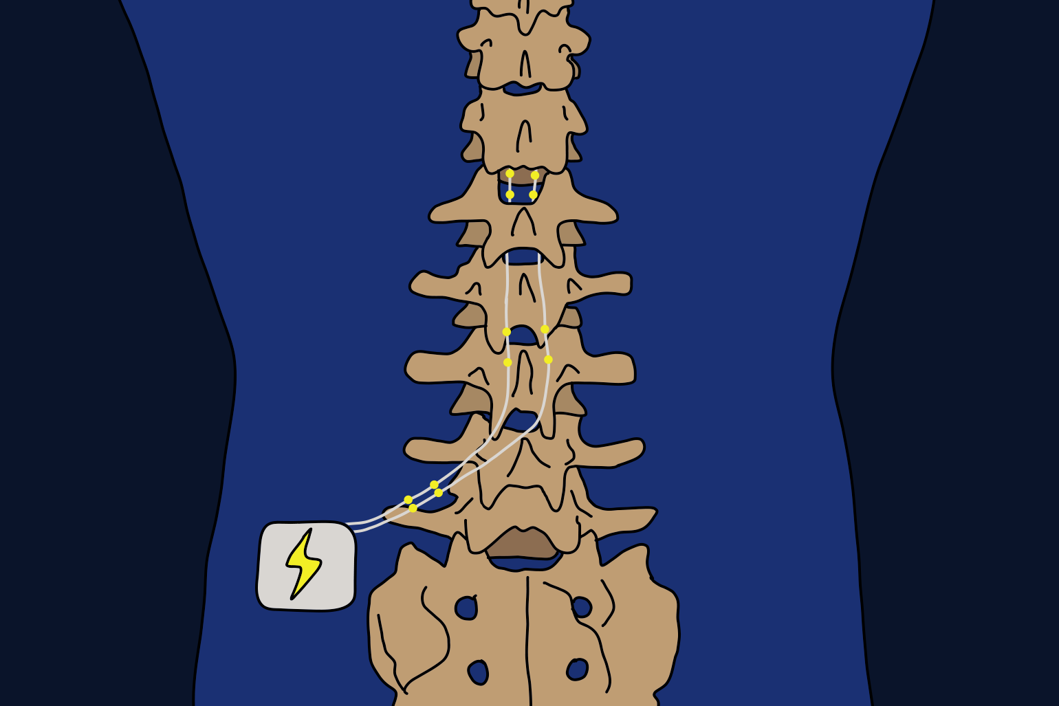
**Kobe University's** groundbreaking study promises a new way to predict the success of spinal cord stimulation surgery—a risky procedure designed for chronic pain sufferers. This surgery involves implanting leads into the spine to stimulate the spinal cord, aiming to alleviate persistent pain. Traditionally, its effectiveness is gauged through trial runs, which are invasive and sometimes risky. To counter this, researchers used **functional magnetic resonance imaging (fMRI)** to detect brain patterns predicting a patient's responsiveness to the surgery. **UENO Kyohei**, an anesthesiologist at Kobe University, explained their findings: the **weaker connectivity between the default mode network and the salience network**, the better patients responded to the treatment. This connectivity is pivotal, as these networks play roles in self-related thoughts, chronic pain, attention, and response to stimuli. Their study, featuring 29 patients with varied chronic pain forms, was published in the British Journal of Anaesthesia. It highlighted that a **10-minute resting state fMRI scan** might serve as a reliable predictor, possibly reducing the burden on patients and healthcare providers. Although diverse patient conditions initially appeared challenging for accurate assessment, the researchers are optimistic about refining predictive capabilities with further studies, aiming to personalize spinal cord stimulation therapy. This research received funding from the **Japan Society for the Promotion of Science** and involved collaboration with Ritsumeikan University experts.