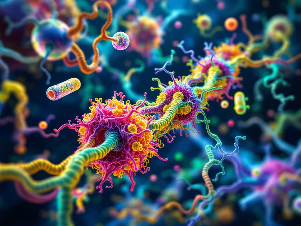
Unveiling New Details in Synaptic Vesicle Architecture with Cryo-Electron Tomography
**Researchers from the Max Delbrück Center**, led by Uljana Kravčenko in Professor Misha Kudryashev's lab, have **advanced our understanding of synaptic vesicles (SVs)** using cryo-electron tomography. This technique allowed for the visualization of SVs in three dimensions, shedding light on important protein interactions and SV recycling mechanisms. **Synaptic vesicles play a critical role in neurotransmission**, storing and releasing neurotransmitters like dopamine and serotonin at the presynaptic terminals of neurons. The study confirmed the interaction between V-ATPase and synaptophysin, a long-suspected but unvisulaized pairing until now. Additionally, researchers identified the spatial organization of clathrin cages, integral to the recycling of SVs, closer to the cell membrane, suggesting an energy-efficient recycling mechanism. **Cryo-electron tomography stands out** by preserving the native state of vesicles, offering clearer protein organization insights compared to other imaging techniques. The findings have potential implications for understanding neurological disorders involving synaptic vesicle dysfunction and may pave the way for diagnostic and treatment advancements.